The X-ray Microscope at ASTRID - Projects
The Aarhus XM is a multi-user facility operating for periods of three months, twice a year. It is used for many different scientific investigations, in such diverse areas as:
Algae
 |
The blue-green algae play an important role in nutrient
recycling in the fresh water environment. Fluctuations in species
composition and population densities can have far reaching effects, with
sporadic toxic algal blooms causing extensive destruction of ecosystems
resulting in the death of many higher organisms, such as fish.
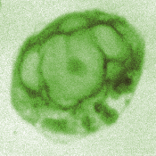
Samornmate Jearanaikoon (better known as Noi) is a PhD student researching structural changes on a variety of blue-green algae, with particular interest in the effects of nutrient conditions, such as phosphorus and nitrogen. XM image of Chlorella (diameter = ~2 mm). |
Human spermatozoa
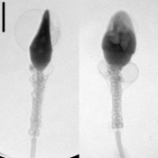
Human spermatozoa at different stages in the maturing process were studied in collaboration with the Department of Medical Microbiology & Immunology, University of Aarhus. We observed by XM that the sperm underwent distinct morphological changes when exposed to capacitating conditions. These morphological changes had not been reported before. Human Reproduction 14, 880-884 (1999).
 |
In a collaborative research project with the Department of
Obstetrics & Gynaecology, University of Manchester, UK, we identified for
the first time mid-piece associated vesicles on human sperm from normozoospermic semen. We quantified the frequency of occurrence of these
membrane changes and found that the incidence is donor specific. Medical
Science Research 23, 663-667 (1998).
XM images of human sperm in a 3-5 mm liquid layer (bar = 2 µm). |
The XM & Andrology Group, was established with the clear objective to find out how the vesicles are generated, how they respond to their environment and what part they play in fertilisation.
We have investigated the osmotic response of mid-piece vesicles on human spermatozoa. Light microscopy, transmission X-ray microscopy and computer-aided semen analysis were used to investigate spermatozoa in normozoospermic semen from healthy donors, separated from semen and suspended in hyper- or hypo-osmotic solutions. We have found that mid-piece vesicles are ubiquitous and distinct from cytoplasmic droplets. They respond to osmolality changes of the surrounding medium. The presence of a mid-piece vesicle can reduce motility but not survival in cervical mucus. Therefore, they should not be considered detrimental to spermatozoa function Human Reproduction 17 (2), 375-382 (2002).
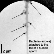 |
Mycoplasma genitalium is a human pathogen causing
urogenital diseases including pelvic inflammatory disease, cervicitis,
endometritis and infertility. It is thought to be sexually transmitted and
is fast becoming the new Chlamydia. Spermatozoa are a possible vector for
the transmission of the bacteria. Helle Friis Svenstrup is researching the
attachment of Mycoplasma genitalium to human spermatozoa by a variety of
imaging techniques, including XM. Human Reproduction 18 (10), 2103-2109
(2003).
More XM images of human sperm. |
Plant photosynthetic membranes
 |
Dr. Dorthe Posselt and Jens Kai Holm, Department of
Mathematics and Physics; Dr.Gyõzõ Garab and Dr. László Kovács, Institute of
Plant Biology, Biological Research Center, Szeged, Hungary are presently
investigating the structure of various model systems related to the
chloroplast thylakoid membrane system using scattering techniques
(small-angle X-ray (SAXS) and small-angle neutron scattering (SANS) probing
length scales from ~1-100nm) as well as different spectroscopic methods,
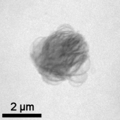
mainly circular dichroism (CD) and chlorophyll fluorescence. X-ray
microscopy with a resolution of 29 nm on these model systems is highly
valuable in supplying additional structural information to the understanding
of these complex systems.
XM image of a thylakoid |
Iron-precipitating bacteria/Iron sludge
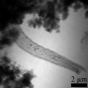 |
Research leader Erik Søgaard, University of Ålborg. There
are high levels of iron in the ground water around Esbjerg, Denmark. The
iron has to be removed before distribution for drinking water. The
biological treatment of drinking water is more efficient than chemical
treatment. We have characterised the morphology of the different types of
precipitated sludge and determined the kinds of bacteria involved in the
oxidation of iron. Water Research 34 (10), 2675-2682 (2000). Applied
Geochemistry 16 (9-10), 1129-1137 (2001) XM image of a sheath deposited by an iron-precipitating bacterium. |
Protozoa and metal toxicity
 |
At ISA, we have used the flagellated protozoon Chilomonas
paramecium, commonly found in polluted water, as a cellular model in the
study of metal toxicity. Europ. J. Protistol., 34 51-57 (1998). Cell Bio.
Int. 25 (6), 521-530 (2001).
We have combined different microscopy techniques (conventional light, confocal fluorescence and X-ray) to determine structural changes induced on exposure to free metal ions, e.g. aluminium, copper, lead and zinc. Elevated levels of free metal ions in the external environment can be toxic to the cell. Using XM we had the unique chance to get high-resolution images of thick specimens in solution. Artefacts caused by dehydration of the sample during other microscopy preparation protocols were eliminated because the cells were in solution throughout imaging. XM image of a whole glutaraldehyde-fixed Chilomonas paramecium still in solution. The cell was previously exposed to elevated levels of lead (bar = 2 µm). |
Last Modified 04 January 2013