The X-ray Microscope at ASTRID
The X-ray microscope was built in the early 1990s, one of the first beam lines to be added to ASTRID. For many years the microscope was used with great success in many research projects, for example looking at human spermatozoa at different stages in the maturing process at the micron scale. While the X-ray microscope is still at ISA, it is not currently in use and there are no plans to move the beam line to the new ring, ASTRID2.
Microscopy in the soft X-ray region is done in a similar way as microscopy with visible light or electrons. In contrast to these techniques the properties of the interaction of X-rays with matter and the wavelength of the X-rays allow a resolution below 25 nm for the observation of objects with a thickness of up to 10 micrometer. In particular, objects in an aqueous medium can be investigated. X-rays produced in a bending magnet of the ASTRID synchrotron are focused by a condenser lens into the object region. An objective lens forms a magnified image of the object onto a CCD detector.
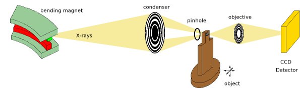
Schematic diagram of the Aarhus X-ray microscope
In X-ray microscopy the optics cannot operate by using the effects of refraction or reflection. Fresnel zone plates based on diffraction are employed instead. These circular gratings have a varying line density. With an increasing distance from the centre the gratings become smaller and smaller, therefore producing an increasing angle of diffraction. If the width of the gratings follows a certain rule, an incoming plane wave is focused into different focal points connected to the different orders of diffraction. For a single order the properties of a zone plate are comparable to that of a lens based on refraction.
Last Modified 30 August 2016