The CD 1 beamline at ASTRID
A new Ultra Violet beamline was commisioned in June 2007 at ISA. The beamline, called CD1, is optimized for and dedicated to the use for Synchrotron Radiation Circular Dichroism (SRCD). It is optimized to operate in the wavelength range of 115-350 nm, and can be used up to a wavelength of 700 nm.
The ASTRID SRCD facility was formerly based on the UV1 beamline and was thus sharing the time with many other UV and VUV experiments. The success of the UV1 beamline as a facility was clear from the number of applications ASTRID receives from both Danish and other European scientists. We could far from accommodate all the users we would like to. In addition, it was very difficult for us to accommodate experiments on a short term notice, which is a problem for the type of technique that CD is: fast and easy. Fast because the data acquisition time typically is less than one hour for a sample, and easy because there is no significant sample preparation. The aim of the CD1 beamline is to establish and run a dedicated SRCD facility, offering quick access to SRCD. This was done by building a UV-VUV beamline (CD1) fully optimized for SRCD on ASTRID.
The technical details of the beamline can be found on the CD1 optical specification page.
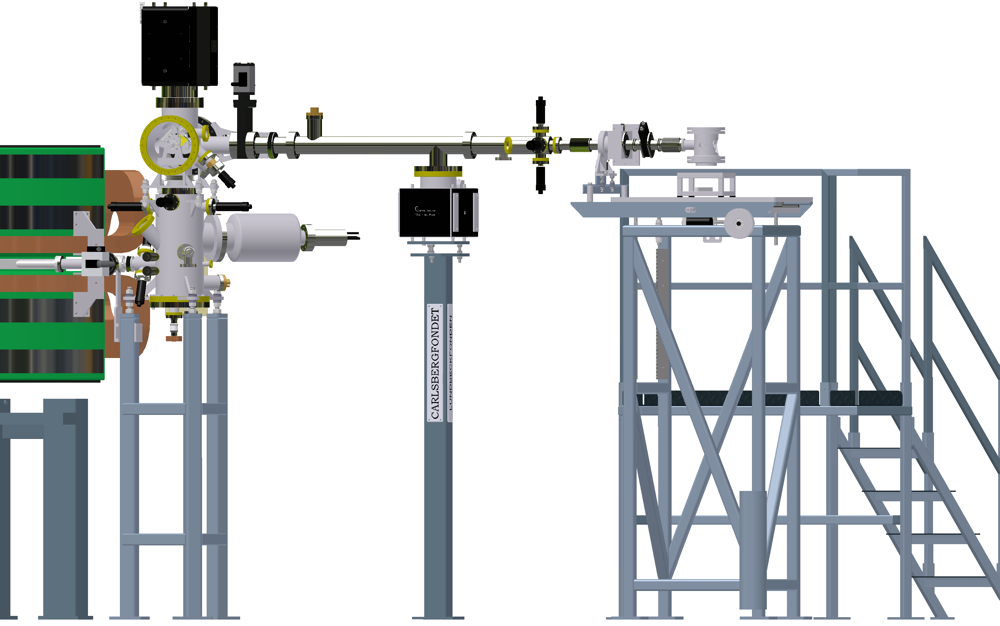
Drawing of the CD1 beamline showing the beam path inside the vacuum vessel.
Both the mirror and the grating can be seen.

Drawing of the CD1 beamline.
Synchrotron Radiation Circular Dichroism.
SRCD spectroscopy offers significant improvements to the well-established method of conventional circular dichroism (cCD) spectroscopy. It takes advantage of the high photon flux available from synchrotron sources over a wide range of wavelengths, which results in higher signal-to-noise ratios and enables the collection of lower wavelength data than possible with cCD spectrometers.
The wavelength range of CD1 (115 to 350 nm) is a biologically important wavelength region, and the high degree of linear polarisation of the radiation, and the low level of scattered light from CD1 makes the beamline well suited for Synchrotron Radiation Circular Dichroism (SRCD) spectroscopy of optically active macromolecules.
Circular Dichroism (CD) spectroscopy measures the difference in absorption between left and right handed circularly polarized light in chiral molecules. It is an established biophysical method probing the secondary structure (e.g. helices, beta-sheets, turns etc.) of peptides, proteins and nucleic acids which have distinct CD bands in the far-UV and VUV.

The principle behind CD spectroscopy. The light from UV1 is passed into a Photo Elastic Modulator (PEM) which converts the linear polarized light into alternating left and right
handed polarized light. The two polarizations are differently absorbed, and the difference in absorption is
detected with a Photo Multiplier Tube (PMT)
Secondary structures give rise to characteristic far UV and VUV CD spectra. A CD spectrum can be considered to arise from the weighted summed components of secondary structure that comprise a protein. Studies have shown that the information content in these data (i.e. how many different types of secondary structures can accurately be distinguished) is highly dependent on the wavelength range of the spectrum. Using data between 200 and 260 nm, the information content is two, rising to three if data down to 190 nm are used. If the data extend down to 178 nm, the value is five, and by taking spectra to 160 nm the information content rises to about eight. Thus the additional wavelength range offered by SRCD greatly enhances the number of secondary structure types that can be distinguished in a CD spectrum.
More details of the present SRCD facility at the UV1 beamline at ASTRID can be found in the SRCD folder.
How to get the best SRCD data.
In any CD experiment in general and especially for a SRCD experiment great care in the choice of buffers and salts should be taken. Chloride for example is one of the ions which should be avoided due to the ions high Far-UV/VUV absorption. This means that e.g. Tris buffers which are pH regulated with HCl must be avoided, and NaCl salt should be substituted with NaF whenever possible. A few guide lines are given in the list below.
- Lowest wavelength data are obtained using short pathlength cells. We have cells with path lengths down to about 10mu.
- If possible, use pure deionized water as a solvent.
- Phosphate buffers are well suited. Please keep concentration below 20mM.
- NaCl salts must be avoided. If possible use NaF instead to obtain ionic strength.
Miles, A.J. and Wallace, B.A. "Synchrotron Radiation Circular Dichroism Spectroscopy of Proteins and Applications in Structural and Functional Genomics". Chem. Soc. Reviews 35 (2006) 39-51.
A general review on how to do CD can be found in:
Kelly, S.M.; Jess, T.J.; Price, N.C. "How to study proteins by circular dichroism" Biochimica et Biophysica Acta 1751 (2005) 119 – 139.
Contact information:
For further information on and access to the CD1 beamline please contact:
Beamline Scientist
Last Modified 15 June 2009


