The X-ray Microscope at ASTRID - Applications
The Aarhus X-ray microscope operates mainly in the so called water window at a wavelength between 2.3 and 4.4 nm. In this region there is a natural contrast between Oxygen and Carbon. For example, the photoelectric absorption of X-rays in water is an order of magnitude lower than in organic material. This contrast makes the microscope ideal for studying biological material. If necessary gold labelling techniques gives additional contrast.
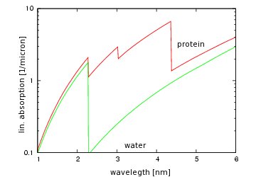
Absorption of different materials in the soft X-ray region
A much higher resolution can be obtained in the X-ray microscope (<30 nm) than in a visible-light microscope (~200 nm). This is due to the shorter wavelength of soft X-rays and the performance of the optics. However, the simplicity of visible-light microscopy sample preparation is not lost. Many of the restrictive sample preparations imposed by EM are eliminated. In X-ray microscopy, the sample DOES NOT have to be thin (i.e. below 1 µm), chemically fixed, stained for contrast or dried. Limitations arise from the thickness of the sample (in generel below 10 micron) and under certain conditions from the object stability during exposure.
Two modes of operation are available. At room temperature imaging, samples are trapped between 2 silicon wafers with transmissive walls thinned to 150 nm in the central part, and then mounted in a special target chamber. Imaging is done at atmospheric pressure. When the object stability becomes an issue cryo imaging is used. Aqueous samples prepared on grids are plunge frozen in liquid ethane. Imaging is then done under vacuum conditions.
Last Modified 04 January 2013